Try to defend a bustling city with the same impenetrable brick wall all year round. Certain elements would be excluded, sure, but so would food, information, and help! Living cells have this exact problem to solve. While a barrier is absolutely necessary to keep some cellular contents from escaping, cells are so dynamic that their membrane needs flexibility for food to get in, waste to get out, and messages between cells to be delivered.
This balance is explained by the fluid mosaic model, one of the most important concepts in modern biology. Rather than describing the cell membrane as a fixed barrier, the model shows it as a dynamic structure in which lipids and proteins move within a flexible membrane. Understanding the fluid mosaic model helps explain how cells survive, communicate, grow, and respond to changes in their environment.
Through this guide you’ll be shown the model the science came up with, what the cell membrane’s ingredients are, why the model is structured the way it is and what that science can be used for in biology, medicine and science research.
Understanding the Foundation
Before exploring the fluid mosaic model itself, it is helpful to understand the structure it describes—the cell membrane.
Plasma (cell) membrane. Within each living cell you can find a delicate structure called a plasma membrane or cell membrane which is present around it separating it from the exterior world. While it is a few nanometers in thickness the membrane has some vital tasks it fulfills within each living cell – such as guarding the cell, managing movement, enabling cellular communication and maintaining internal environment stability.
Without an effective membrane, cells would be unable to control what enters or leaves, making life impossible.
What Is the Fluid Mosaic Model?
The fluid mosaic model is a scientific model that explains the structure and behavior of the cell membrane.
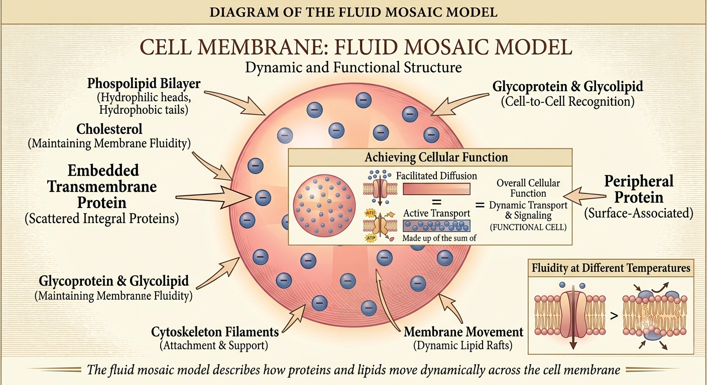
It suggests the membrane structure is based around two layers of lipids, called a phospholipid bilayer, where some parts are proteins, others cholesterol, and others carbohydrates are inserted or associated with it, and where the different elements are in constant motion; they move laterally within the membrane and so the structure and arrangement is dynamic rather than fixed.
The word ‘fluid’ refers to the fact that the components of the membrane are constantly moving. The word ‘mosaic’ refers to the mosaic of various types of lipids, proteins, and carbohydrate throughout the membrane-just like various colored tile work to compose a mosaic. This model accounts for the flexibility of the cell membrane and its relative rigidity to provide a barrier.
Historical Background
Even prior to the invention of fluid mosaic model, scientists had various proposals about structure of the membrane which could not account for many observations. The fluid mosaic model: In 1972 biologists S. Jonathan Singer and Garth L. Nicolson published their seminal work on the fluid mosaic model of the structure of cell membranes, which culminated years of experimental observation involving a combination of microscopy, biochemistry, and thermodynamics.
These developments, driven by advances in imaging technology and molecular biology, continue to build on our understanding. Scientists are learning that certain regions of the membrane may not be as haphazardly arranged as we first thought, but the fluid mosaic model still provides the conceptual framework with which we all learn about what the cell membrane does and how it’s constructed.
Why the Fluid Mosaic Model Matters
If you take a quick look, it’s easy to confuse with a drawing in some biological schoolbook. On the other hand it actually reveals processes happening every day, all the time, in every organism existing.
The fluid mosaic model helps scientists understand:
- How nutrients enter cells.
- How waste products leave cells.
- How nerve cells transmit signals.
- How hormones communicate with target cells.
- How the immune system recognizes foreign substances.
- How certain diseases affect membrane function.
- How medicines interact with cells.
Without this model, many biological processes would be much harder to explain.
Breaking Down the Cell Membrane
To understand the fluid mosaic model, it helps to examine each major component individually.
Phospholipid Bilayer
The phospholipid bilayer forms the basic framework of the membrane.
Each phospholipid molecule contains two different regions:
- A hydrophilic (water-attracting) head
- Two hydrophobic (water-repelling) fatty acid tails
Since our environment is aqueous (watery), lipids self assemble into two layers. This allows both of the hydrophilic heads of the lipids to expose toward the water on each side of the membrane. Alternatively, the tail parts of the phospholipids face away from the water. This forms an ever so stable, fluid membrane that divides cells.
If you imagine the phospholipid bilayer being organized like two matchstick sticks that were aligned head-to-head, with the heads being on the outside and the wood portions going in the center then you know what we’re talking about. This means we don’t destroy the integrity of the cell, while keeping mobility possible.
Membrane Proteins
Proteins are scattered throughout the phospholipid bilayer, giving the membrane much of its functionality.
Different proteins perform different jobs.
Some transport molecules across the membrane.
Others receive chemical signals.
Some help neighboring cells recognize one another.
Still others anchor the membrane to internal cellular structures.
Membrane proteins generally fall into two categories.
Integral Proteins
Integral proteins extend partially or completely through the membrane.
They often function as:
- Transport channels
- Carrier proteins
- Receptors
- Enzymes
Because they pass through the phospholipid bilayer, they can interact with both the inside and outside of the cell.
Peripheral Proteins
Peripheral proteins attach only to the inner or outer surface of the membrane.
They usually assist with:
- Cell signaling
- Structural support
- Communication
- Cytoskeleton attachment
Although they do not penetrate the membrane, they remain essential for many cellular activities.
Cholesterol
Another important component of an animal cell membrane is cholesterol. While cholesterol has a stigma of health and cholesterol problems attached to it, it also plays a valuable biological role within cell membranes.Testerol also prevents cells from packing too close to one another when the temperature is low and does not let there be too much fluidity if it’s hot
Through this mechanism cholesterol functions like a thermostat, allowing cell membranes to maintain ideal flexibility as environmental changes occur.
Carbohydrates
Others membrane proteins and lipids may have chains of carbohydrates that extend to the exterior surface.
These molecules form structures known as:
- Glycoproteins
- Glycolipids
Carbohydrates contribute to:
- Cell recognition
- Immune responses
- Cell communication
- Tissue organization
For example, immune cells often distinguish the body’s own cells from foreign invaders by recognizing these surface carbohydrate markers.
How the Different Parts Work Together
One of the strengths of the fluid mosaic model is that it explains how different membrane components cooperate rather than functioning independently.
Imagine a busy harbor.
The water that all activity rests in is the lipid bilayer, acting as sort of a support system. Proteins serve as dock workers and cargo moving, a kind of gated entry, or individual laborers at work; cholesterol allows the choppy seas to stay smooth amidst various temperature swings. Carbohydrates can be viewed as identifying flags that keep the ship in the right harbor; all elements fulfill specific individual jobs while cohesively establishing a very complex system in a rather tight package.
Membrane Fluidity Explained
The concept of membrane fluidity is central to the fluid mosaic model.
Fluidity refers to the ability of phospholipids and many proteins to move sideways within the membrane.
This movement is important because it allows cells to:
- Repair damaged membrane areas.
- Grow during cell division.
- Transport molecules efficiently.
- Respond to environmental changes.
- Communicate with neighboring cells.
- Rearrange membrane proteins when needed.
If the membrane were completely rigid, these essential processes would become extremely difficult or impossible.
Factors That Influence Membrane Fluidity
Although membranes are fluid, their flexibility is not constant.
Several factors influence membrane behavior.
Temperature
Higher temperatures generally increase membrane fluidity, while lower temperatures reduce molecular movement.
Fatty Acid Composition
Phospholipids containing unsaturated fatty acids create more space between molecules, increasing flexibility.
Saturated fatty acids pack more closely together, making the membrane less fluid.
Cholesterol Levels
As discussed earlier, cholesterol acts as a stabilizer, preventing extreme changes in membrane fluidity caused by temperature fluctuations.
These factors work together to ensure the membrane remains functional under different physiological conditions.
Fluid Mosaic Model: A Complete Educational Guide
Selective Permeability: The Membrane’s Most Important Property
One of the greatest strengths of the fluid mosaic model is its ability to explain selective permeability.
Simply put,Selective Permeability is theability ofa cell membraneto permitthe passageofsome substances butinhibit thepassage of others. Rather than bein adoorway orwall, the membraneis morelike a well-trained checkpoint guard. The membraneinspects eachtype of Substance that comes its way foritssize, chargand compositionbeforeallowingoit o notalloit to enter.
For example:
- Oxygen and carbon dioxide can move relatively easily through the phospholipid bilayer.
- Water moves primarily through specialized protein channels called aquaporins, although a small amount can diffuse directly.
- Large molecules such as glucose usually require transport proteins.
- Charged ions like sodium (Na⁺), potassium (K⁺), and calcium (Ca²⁺) cannot cross the lipid bilayer freely and depend on ion channels or pumps.
This selective control helps maintain homeostasis, the stable internal environment required for normal cellular function.
How Materials Move Across the Membrane
The fluid mosaic model also explains why cells use different transport mechanisms depending on the type of molecule being moved.
Simple Diffusion
Simple diffusion is the movement of molecules from an area of higher concentration to an area of lower concentration without requiring energy.
Examples include:
- Oxygen entering cells
- Carbon dioxide leaving cells
Because these molecules are small and nonpolar, they pass directly through the phospholipid bilayer.
Facilitated Diffusion
Some molecules cannot pass through the membrane independently.
Instead, they move through specialized membrane proteins that act like tunnels or gateways.
Examples include:
- Glucose
- Certain amino acids
- Some ions
Facilitated diffusion still moves substances down their concentration gradient and does not require cellular energy (ATP).
Osmosis
Osmosis is the diffusion of water across a selectively permeable membrane.
Water naturally moves toward areas with a higher concentration of dissolved substances until equilibrium is reached.
Osmosis helps regulate:
- Cell volume
- Water balance
- Nutrient distribution
- Waste removal
Understanding osmosis is essential in biology because even slight changes in water balance can affect cell survival.
Active Transport
Sometimes cells must move substances against their concentration gradient.
This process requires energy, usually supplied by ATP.
Active transport is essential for maintaining proper concentrations of:
- Sodium ions
- Potassium ions
- Calcium ions
- Hydrogen ions
A well-known example is the sodium-potassium pump, which helps nerve cells transmit electrical signals and supports many other physiological processes.
Cell Communication and the Fluid Mosaic Model
Constant Communication between the Environment and Cells Receptors (part of Membrane Proteins) help with information exchange between the surrounding environment and the inside of the cell. Signaling molecules bind to receptors triggering a response. Signaling molecules include hormones,neuromitters, andgrowth factors
These responses may include:
- Activating genes
- Starting enzyme reactions
- Stimulating cell growth
- Coordinating immune responses
- Regulating metabolism
The fluid nature of the membrane allows receptor proteins to move and interact efficiently with other membrane components.
Cell Recognition and Immune Function
Carbohydrates attached to proteins and lipids help cells identify one another.
These carbohydrate chains act like biological identification tags.
They help the immune system distinguish:
- Healthy body cells
- Invading bacteria
- Viruses
- Transplanted tissues
For example, blood group antigens in the ABO blood typing system are carbohydrate markers found on the surface of red blood cells.
Without these recognition molecules, immune responses would become much less accurate.
Real-World Applications of the Fluid Mosaic Model
Although the fluid mosaic model is taught in biology classes, its applications extend far beyond the classroom.
Medicine
Many medications interact directly with membrane proteins.
Examples include drugs that target:
- Hormone receptors
- Ion channels
- Transport proteins
- Enzymes embedded in the membrane
Understanding membrane structure helps researchers design more effective treatments.
Biotechnology
Biotechnology applies membrane biology to advance commercial innovations—from targeted drug delivery to biosensors—many of which drive the health-tech sectors covered on Tech Coore.
- Drug delivery systems
- Artificial membranes
- Biosensors
- Cell culture techniques
- Gene therapy methods
Scientists continue to apply membrane biology in developing innovative medical technologies.
Neuroscience
Nerve cells and their electrical activity The selective transport of ions across a neuron’s cell membrane must be tightly managed. As such, thefluid mosaicmodelofthe membrane provides an explanation for how neurons transmit an electrical signal along their cells and throughout the nervous system by passing the signal onto other cells or utilizing and processing signals.
Infectious Disease Research
Many viruses begin infection by attaching to receptor proteins on the cell membrane.
Learning how these interactions occur helps scientists develop vaccines, antiviral medicines, and strategies to reduce infection.
Common Misconceptions
Understanding the fluid mosaic model also means recognizing what it does not say.
Misconception 1: The Membrane Is Completely Liquid
The membrane is fluid, but it is not a free-flowing liquid. Other proteins tethered to the cytoskeleton or to the extracellular matrix cannot move far from their positions.
Misconception 2: Every Molecule Moves Freely
Although many phospholipids and proteins move laterally, movement is regulated. Certain membrane regions are more organized than others.
Misconception 3: Only Phospholipids Matter
Phospholipids form the membrane’s framework, but proteins, cholesterol, and carbohydrates are equally important for membrane function.
Misconception 4: The Fluid Mosaic Model Is Outdated
While scientists have refined the original model by discovering specialized membrane domains and additional complexities, the fluid mosaic model remains the accepted foundation for understanding biological membranes.
Modern Refinements to the Model
Microscope and molecular biology has demonstrated more dynamic than first envisioned that membranes is. Researchers found regions, lipid rafts in a specialized, temporarily gathered molecules of proteins in this or specific lipoids. Microdomains seem involved in signaling, membrane processes in immune function.
Furthermore scientists had shown that the cytoskeleton could act on membranes and reduce mobility on some membrane proteins. The new ideas do not contradict the existing ones but add detail and indicate that membranes are fluid structures but they are locally structured to control molecule behavior.
Tips for Learning the Fluid Mosaic Model
Students often find this topic easier when they focus on understanding rather than memorization.
A few strategies can help:
- Visualize the membrane as a flexible phospholipid sea with proteins floating within it.
- Find out each one alone first, then see how they work with other one together.
- Connect membrane transport processes with selective permeability.
- Practice drawing and labeling the membrane structure.
- Relate membrane functions to real biological examples, such as nerve signaling or hormone action.
If you know why it is structured in this way, then it will stick around much easier for you to remember.
Conclusion
The fluid mosaic model transformed our understanding of cell biology by showing that the cell membrane is neither rigid nor randomly organized. Instead, it is a flexible phospholipid bilayer containing proteins, cholesterol, and carbohydrates that work together to regulate transport, communication, recognition, and stability.
While a modern version the model offers additional insights in to cell functions, the fundamental premises set by the original fluid mosaic model are essential in biological study today. It provides us with understanding as to how cells can achieve homeostasis, regulate substance transfer, inter-cellular communication and regulate environmental interactions.
From a student preparing for an exam to someone starting a biology class or just wanting to know what the biological world has to offer; you need a good grasp of the fluid mosaic model to begin to learn many other areas in the study of life.
Frequently Asked Questions
1. What is the fluid mosaic model in simple terms?
The fluid mosaic model describes the cell membrane as a flexible phospholipid bilayer containing proteins, cholesterol, and carbohydrates that move within the membrane and perform specialized functions.
2. Why is it called the “fluid mosaic model”?
The membrane is called fluid because many of its molecules can move sideways within the bilayer. It is called mosaic because it contains a diverse arrangement of lipids, proteins, and carbohydrates.
3. Who proposed the fluid mosaic model?
The model was first described in 1972 by S. Jonathan Singer and Garth L. Nicolson as an explanation for the structure and function of biological membranes.
4. What are the basic parts of the cell membrane?
The cell membrane consists primarily of a phospholipid bilayer, membrane proteins, cholesterol (in animal cells), and carbohydrates attached to proteins and lipids.
5. How does the fluid mosaic model explain selective permeability?
The model shows that the phospholipid bilayer blocks many substances, while specialized membrane proteins allow selected molecules and ions to move into or out of the cell.
6. Is the fluid mosaic model still accepted today?
Yes. WhileScientists developed the fluidity and protein sandwich model later on, to include additional facts about cell membranes such as rafts and microdomains, it is still the base understanding of the cell membrane.